
具体描述
商品参数
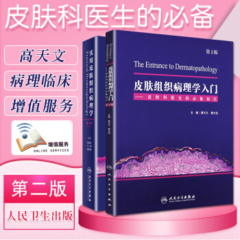
皮肤组织病理学入门 9787117262217
实用皮肤组织病理学 9787117253420
内容介绍
皮肤组织病理学入门
正常皮肤的组织结构和炎症细胞 (structure of normal skin and inflammatory cells)
1.1 皮肤的基本组织结构 (structure of skin)
1.1.1 正常皮肤组织(normal skin)
1.1.2 表皮(epidermis)
1.1.3 真皮(dermis)
1.1.4 皮下组织(subcutaneous tissue)
1.1.5 皮肤附属器(skin appendages)
1.2 特殊部位的组织结构(normal tissue structure of special anatomic site)
1.2.1 头皮(scalp)
1.2.2 口唇(lip)
1.2.3 眼睑(blephara)
1.2.4 生殖器部位(genital organ)
1.2.5 掌跖(vola)
1.2.6 乳房(breast)
1.2.7 耳廓(ala auris)
1.2.8 甲(nail)
1.3炎症细胞(inflammatory cells)
1.3.1 淋巴细胞(lymphocyte)
1.3.2 组织细胞(histocyte)
1.3.3 多核巨细胞(multinuclear giant cell)
1.3.4 浆细胞(plasma cell)
1.3.5 中性粒细胞(neutrophil)
1.3.6 嗜酸性粒细胞(eosinophil)
1.3.7 肥大细胞(mast cell)
2 皮肤病的基本病理变化(basic pathological change in skin)
2.1 表皮病变(terms of the disorders in epidermis)
2.1.1 角化过度(hyperkeratosis)
2.1.2 角化不全(parakeratosis)
2.1.3 角化不良(dyskeratosis)
2.1.4 颗粒层增厚(hyperkeratosis)
2.1.5 颗粒层减少(hypokeratosis)
2.1.6 棘层肥厚(acanthosis)
2.1.7 假上皮瘤样增生(pseudoepitheliomatous hyperplasia)
2.1.8 表皮萎缩(epidermal atrophy)
2.1.9 表皮水肿(epidermal edema)
2.1.10 嗜酸性海绵形成(eosinophilic spongiosis)
2.1.11 棘层松解(acantholysis)
2.1.12 绒毛(villi)
2.1.13 基底细胞液化变性(liquifaction degeneration of basal cells)及色素失禁(incontinence of pigment)
2.1.14 空泡细胞(koilocyte)
2.1.15 水疱(blister)和大疱(bulla)
2.1.16 脓疱(pustule)
2.1.17 嗜酸性微脓肿(eosinophilic microabscesses)
2.1.18 Pautrier微脓肿(Pautrier microabscesses)
2.1.19 细胞外渗(exocytosis)
2.1.20 亲表皮性(epidermotropism)
2.1.21 表皮松解性角化过度(epidermolytic hyperkeratosis)
2.1.22 痂(crust)
2.1.23 色素增多(hyperpigmentation)
2.1.24 色素减少(hypopigmentation)
2.1.25 色素传输障碍(melanin transfer blockade)
2.1.26 毛囊角栓(follicular plug)
2.1.27 鳞状涡(squamous addy)
2.1.28 角囊肿(horn cyst)
2.1.29 外毛根鞘角化(trichilemmal keratinization)
2.2 真皮病变(terms of the disorders in dermis)
2.2.1 乳头状瘤样增生(papillomatosis)
2.2.2 境界带(grenz zone)
2.2.3 收缩间隙(retraction Space)
2.2.4 透明变性(hyaline degeneration)
2.2.5 胶样变性(colloid degeneration)
2.2.6 嗜碱性变性(basophilic degeneration)
2.2.7 淀粉样变性(amyloid degeneration)
2.2.8 纤维蛋白样变性(fibrinoid degeneration)
2.2.9 黏液变性(mucinous degeneration)
2.2.10 弹力纤维变性(degeneration of elastic fibers)
2.2.11 渐进性坏死(necrobiosis)
2.2.12 色素沉积(pigment deposition)
2.2.13 脂质沉积(fatty deposition)
2.2.14 钙沉积(calcinosis)
2.2.15 血管闭塞(vascular obliteration)
2.2.16 血栓形成(thrombosis)
2.2.17 肉芽组织(granulation tissue)
2.2.18 肉芽肿(granuloma)
2.2.19 彩球状(pompon-like)
2.3 皮下组织病变(terms of the disorders in subcutaneous tissue)
2.3.1 增生性萎缩(proliferation atrophy or wucher atrophy)
2.3.2 脂膜炎(panniculitis)
2.3.3 脂肪细胞坏死(fat cell necrosis)
2.3.4 Miescher放射状肉芽肿(Miescher radial granuloma)
2.4 普通病理改变(terms in general histopathology)
2.4.1 坏死(necrosis)
2.4.2 凋亡(apoptosis)
2.4.3 核固缩(karyopyknosis)
2.4.4 核碎裂(karyorrhexis)
2.4.5 核溶解(karyolysis)
2.4.6 萎缩(atrophy)
2.4.7 间变(anaplasia)
2.4.8 异型性(atypia)
2.4.9 错构瘤(hamartoma)
2.4.10 机化(organization)
3特殊染色(characteristic staining)
3.1 组织化学染色(characteristic staining of tissue and cell)
3.1.1 淀粉样物质染色(staining of amyloid substance)
3.1.2 黏液物质染色(staining of mucosubstance)
3.1.3 肥大细胞染色(staining of mast cell)
3.1.4 PAS方法(staining of PAS)
3.1.5 抗酸染色(acid fast stain)
3.1.6 胶原纤维染色(staining of collagen fibers)
3.1.7 弹力纤维染色(staining of elastic fibers)
3.1.8 网织纤维染色(staining of reticular fiber)
3.2 免疫组织化学染色(immunohistochemical stain)
3.2.1上皮来源抗体(epithelial origin antibody)
3.2.2间质和肌性分化抗体(stroma and muscle antibody)
3.2.3血管和淋巴管内皮分化抗体(vascular and lymphatic endothelium antibody
目录
实用皮肤组织病理学
内容包含病理要点、病理鉴别,临床要点、临床鉴别,别名五部分,均为要点,一目了然。病例取自作者单位10余万例的病理数据库,极罕见病例由国内及国际同行提供。以现代病理图片扫描系统采图,每一疾病包含典型、非典型病例,兼顾临床特点、病理特点,选取多个病例取低中高倍图,必要的免疫组织化学图片,全方位介绍一个疾病。
用户评价
第二段: 作为一名多年的皮肤科老医生,我手中也曾阅览过不少皮肤病学相关的书籍,但《皮肤组织病学入门+实用皮肤组织病学》给我带来的惊喜依然不小。这本书的编排设计非常巧妙,它既能满足初学者打牢基础的需求,也为我这样的资深从业者提供了进一步精进的参考。书中的一些罕见病和疑难杂症的组织病理学分析,让我耳目一新,甚至发现了一些我之前可能忽略的细微鉴别点。尤其是关于一些鉴别诊断的环节,作者列举了多种相似病变的组织学特征,并进行了详尽的对比分析,这对于我们在实际工作中遇到模棱两可的病例时,提供了非常有价值的思路。我特别欣赏书中对一些经典病例的深入剖析,它们不仅仅是图片和文字的简单罗列,而是包含了一套完整的诊断思路和鉴别流程,这对于我们提升临床思维能力非常有益。此外,书中的语言风格也比较严谨而不失生动,阅读起来不会感到枯燥乏味,反而能激发起我深入研究的兴趣。总的来说,这是一本值得反复研读,并能在职业生涯中不断带来新收获的专业书籍。
评分第一段: 这本《皮肤组织病学入门+实用皮肤组织病学》果真名不虚传,简直是皮肤科新手入门的“圣经”!我是一名刚开始接触皮肤病学的医学生,一开始面对那些琳琅满目的皮肤病变,简直是一头雾水。看这本书就像是有了航海图,里面的讲解条理清晰,从最基础的皮肤结构、生理功能讲起,然后逐步深入到各种常见皮肤病的组织病理学表现。每一章的配图都非常精美,放大镜下的细胞形态、组织结构都展现得淋漓尽致,让我这个“小白”也能大致辨认出一些关键的病理特征。尤其是那些关于炎症、肿瘤、感染性疾病的章节,作者用通俗易懂的语言解释了复杂的病理过程,还搭配了很多临床病例的组织切片,看完之后,我感觉自己对皮肤病学的理解瞬间提升了好几个档次。这本书最大的优点在于它的“实用性”,它不仅仅是理论的堆砌,更强调了如何将组织病理学知识应用到实际的临床诊断中,这对于我这样需要快速掌握技能的初学者来说,简直是雪中送炭。我还会反复翻阅这本书,尤其是那些难懂的部分,相信随着经验的积累,我对书中的内容会有更深刻的体会。
评分第五段: 作为一名皮肤科的独立执业医师,我一直在寻找一本能够帮助我快速查阅、加深理解的参考书。《皮肤组织病学入门+实用皮肤组织病学》无疑是我的不二之选。这本书的编排非常人性化,从基础到临床,层层递进,对于一些复杂的病理学概念,作者都给出了清晰易懂的解释。我尤其欣赏书中提供的“实用”部分,它不仅仅是理论的堆砌,更是将理论知识与临床实践紧密结合,提供了大量的图谱和案例分析,帮助我们快速对照、鉴别。当我遇到一些棘手的病例,或者需要重新审视一些经典病例时,我都会翻阅这本书,它总能给我提供新的视角和启发。书中的图片质量非常高,清晰度和分辨率都非常好,这对于我们进行细微的病理观察至关重要。此外,书中的语言风格既专业又易于理解,避免了过多的生僻术语,让我在阅读时能够更专注于内容的理解。总而言之,这是一本集理论性、实用性、易读性于一体的优秀著作,对于任何从事皮肤科工作的医生来说,都是一本不可或缺的工具书。
评分第三段: 我是一名在皮肤科进修的住院医师,这本书就像是我工作中的“秘密武器”。每次拿到一个病理切片,我都会习惯性地翻阅这本书。它里面的图谱太丰富了,而且标注得非常清晰,什么细胞是正常的,什么细胞是异常的,一眼就能看出个大概。我尤其喜欢书中关于不同类型皮损的组织学特征的归纳总结,比如对于银屑病、湿疹、苔藓样变等,都有非常详细的描述和对比。刚开始的时候,我经常会把一些相似的病变搞混,但有了这本书,我可以对照着图片,找出它们在细胞形态、角质层、棘层、真皮乳头层等方面的细微差别。书中的一些提示和技巧,比如在染色上需要注意什么,在镜下观察时要重点看哪些区域,都非常实用,能帮助我们更快更准确地做出判断。虽然书名听起来有点“学术”,但实际内容一点都不枯燥,反而充满了解决问题的思路和方法,让我感觉自己在学习中不断进步,对皮肤组织病理学的理解也越来越深刻。
评分第四段: 我是一位对皮肤健康有着浓厚兴趣的普通读者,虽然我不是医学专业人士,但我一直对皮肤的奥秘非常着迷。偶然的机会,我看到了这本《皮肤组织病学入门+实用皮肤组织病学》,抱着好奇的心态翻阅了一下。令我惊喜的是,这本书虽然是专业书籍,但对于我这样的大众读者来说,也并非完全无法理解。作者用一种相对易于接受的方式,解释了皮肤的微观世界,让我了解了皮肤的不同层次是如何运作的,以及当皮肤出现问题时,这些微观结构会发生怎样的变化。书中的很多图片都像是一扇扇窗户,让我得以窥见肉眼看不到的细胞和组织,了解皮肤病的根源所在。虽然有些专业术语对我来说还是有些陌生,但我可以通过图片和作者的简单解释,大致领会其意。这本书让我对皮肤有了更深层次的认识,也让我对那些皮肤疾病的治疗有了更科学的理解,不再仅仅停留在表面症状的认识上。它让我明白,皮肤的健康,是无数微小细胞和组织协同工作的结果。
相关图书
本站所有内容均为互联网搜索引擎提供的公开搜索信息,本站不存储任何数据与内容,任何内容与数据均与本站无关,如有需要请联系相关搜索引擎包括但不限于百度,google,bing,sogou 等,本站所有链接都为正版商品购买链接。
© 2026 windowsfront.com All Rights Reserved. 静流书站 版权所有


