
具体描述
商品参数
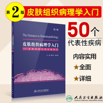
| 皮肤组织病理学入门——皮肤科医生的必备知识(第2版) | ||
| 定价 | 96.00 | |
| 出版社 | 人民卫生出版社 | |
| 版次 | ||
| 出版时间 | ||
| 开本 | ||
| 作者 | ||
| 装帧 | ||
| 页数 | ||
| 字数 | ||
| ISBN编码 | 9787117262217 | |
内容介绍
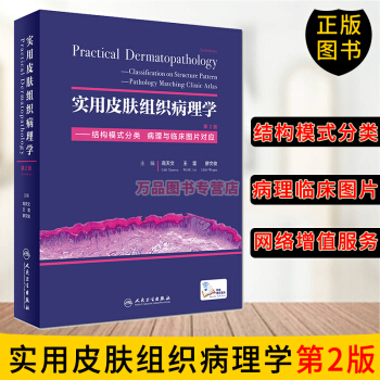
皮肤病理是皮肤科医生的必备知识,不论对其有兴趣与否,皮肤科医生均需学习相关内容。本书适合初学者,可作培训教材之用。另外,本书是已经出版的大型专著《实用皮肤组织病理学》(第2版)的总论部分,需先学习本书再学习《实用皮肤组织病理学》。
本书分四部分,DIYI部分为正常皮肤的组织结构和炎症细胞,主要介绍皮肤的组织发生及结构;第二部分为皮肤病的基本病理变化,介绍了皮肤病的常见病理变化及病理术语;第三部分为特殊染色,简述了皮肤病理取材及常用诊断技术;第四部分为50种代表性的疾病。
初学者只需花半个月左右的时间,在掌握了皮肤病理的基本知识后,掌握ZUI常行皮肤病理检查的50种疾病,为进一步学习《实用皮肤组织病理学》等打好基础。
目录
1 正常皮肤的组织结构和炎症细胞 (structure of normal skin and inflammatory cells)
1.1 皮肤的基本组织结构 (structure of skin)
1.1.1 正常皮肤组织(normal skin)
1.1.2 表皮(epidermis)
1.1.3 真皮(dermis)
1.1.4 皮下组织(subcutaneous tissue)
1.1.5 皮肤附属器(skin appendages)
1.2 特殊部位的组织结构(normal tissue structure of special anatomic site)
1.2.1 头皮(scalp)
1.2.2 口唇(lip)
1.2.3 眼睑(blephara)
1.2.4 生殖器部位(genital organ)
1.2.5 掌跖(vola)
1.2.6 乳房(breast)
1.2.7 耳廓(ala auris)
1.2.8 甲(nail)
1.3炎症细胞(inflammatory cells)
1.3.1 淋巴细胞(lymphocyte)
1.3.2 组织细胞(histocyte)
1.3.3 多核巨细胞(multinuclear giant cell)
1.3.4 浆细胞(plasma cell)
1.3.5 中性粒细胞(neutrophil)
1.3.6 嗜酸性粒细胞(eosinophil)
1.3.7 肥大细胞(mast cell)
2 皮肤病的基本病理变化(basic pathological change in skin)
2.1 表皮病变(terms of the disorders in epidermis)
2.1.1 角化过度(hyperkeratosis)
2.1.2 角化不全(parakeratosis)
2.1.3 角化不良(dyskeratosis)
2.1.4 颗粒层增厚(hyperkeratosis)
2.1.5 颗粒层减少(hypokeratosis)
2.1.6 棘层肥厚(acanthosis)
2.1.7 假上皮瘤样增生(pseudoepitheliomatous hyperplasia)
2.1.8 表皮萎缩(epidermal atrophy)
2.1.9 表皮水肿(epidermal edema)
2.1.10 嗜酸性海绵形成(eosinophilic spongiosis)
2.1.11 棘层松解(acantholysis)
2.1.12 绒毛(villi)
2.1.13 基底细胞液化变性(liquifaction degeneration of basal cells)及色素失禁(incontinence of pigment)
2.1.14 空泡细胞(koilocyte)
2.1.15 水疱(blister)和大疱(bulla)
2.1.16 脓疱(pustule)
2.1.17 嗜酸性微脓肿(eosinophilic microabscesses)
2.1.18 Pautrier微脓肿(Pautrier microabscesses)
2.1.19 细胞外渗(exocytosis)
2.1.20 亲表皮性(epidermotropism)
2.1.21 表皮松解性角化过度(epidermolytic hyperkeratosis)
2.1.22 痂(crust)
2.1.23 色素增多(hyperpigmentation)
2.1.24 色素减少(hypopigmentation)
2.1.25 色素传输障碍(melanin transfer blockade)
2.1.26 毛囊角栓(follicular plug)
2.1.27 鳞状涡(squamous addy)
2.1.28 角囊肿(horn cyst)
2.1.29 外毛根鞘角化(trichilemmal keratinization)
2.2 真皮病变(terms of the disorders in dermis)
2.2.1 乳头状瘤样增生(papillomatosis)
2.2.2 境界带(grenz zone)
2.2.3 收缩间隙(retraction Space)
2.2.4 透明变性(hyaline degeneration)
2.2.5 胶样变性(colloid degeneration)
2.2.6 嗜碱性变性(basophilic degeneration)
2.2.7 淀粉样变性(amyloid degeneration)
2.2.8 纤维蛋白样变性(fibrinoid degeneration)
2.2.9 黏液变性(mucinous degeneration)
2.2.10 弹力纤维变性(degeneration of elastic fibers)
2.2.11 渐进性坏死(necrobiosis)
2.2.12 色素沉积(pigment deposition)
2.2.13 脂质沉积(fatty deposition)
2.2.14 钙沉积(calcinosis)
2.2.15 血管闭塞(vascular obliteration)
2.2.16 血栓形成(thrombosis)
2.2.17 肉芽组织(granulation tissue)
2.2.18 肉芽肿(granuloma)
2.2.19 彩球状(pompon-like)
2.3 皮下组织病变(terms of the disorders in subcutaneous tissue)
2.3.1 增生性萎缩(proliferation atrophy or wucher atrophy)
2.3.2 脂膜炎(panniculitis)
2.3.3 脂肪细胞坏死(fat cell necrosis)
2.3.4 Miescher放射状肉芽肿(Miescher radial granuloma)
2.4 普通病理改变(terms in general histopathology)
2.4.1 坏死(necrosis)
2.4.2 凋亡(apoptosis)
2.4.3 核固缩(karyopyknosis)
2.4.4 核碎裂(karyorrhexis)
2.4.5 核溶解(karyolysis)
2.4.6 萎缩(atrophy)
2.4.7 间变(anaplasia)
2.4.8 异型性(atypia)
2.4.9 错构瘤(hamartoma)
2.4.10 机化(organization)
3特殊染色(characteristic staining)
3.1 组织化学染色(characteristic staining of tissue and cell)
3.1.1 淀粉样物质染色(staining of amyloid substance)
3.1.2 黏液物质染色(staining of mucosubstance)
3.1.3 肥大细胞染色(staining of mast cell)
3.1.4 PAS方法(staining of PAS)
3.1.5 抗酸染色(acid fast stain)
3.1.6 胶原纤维染色(staining of collagen fibers)
3.1.7 弹力纤维染色(staining of elastic fibers)
3.1.8 网织纤维染色(staining of reticular fiber)
3.2 免疫组织化学染色(immunohistochemical stain)
3.2.1上皮来源抗体(epithelial origin antibody)
3.2.2间质和肌性分化抗体(stroma and muscle antibody)
3.2.3血管和淋巴管内皮分化抗体(vascular and lymphatic endothelium antibody)
3.2.4 神经和黑素细胞抗体(nerve and melanocyte antibody)
3.2.5皮肤淋巴增生性疾病相关抗体(antibody for lymphoproliferative disease)
3.2.6组织细胞增生性疾病和肥大细胞相关抗体(histocyte and mast cell antibody)
3.2.7增殖相关抗体(antibody for proliferation)
4代表性疾病(typical entities)
4.1 扁平苔藓(lichen planus)
4.2 多形红斑(erythema multiforme)
4.3 盘状红斑狼疮(discoid lupus erythematosus)
4.4 湿疹(eczema)
4.5 淤滞性皮炎(stasis dermatitis)
4.6 银屑病(psoriasis)
4.7 神经性皮炎(neurodermatitis)
4.8 寻常疣(verruca vulgaris)
4.9 传染性软疣(molluscum contagiosum)
4.10 天疱疮(pemphigus)
4.11 单纯疱疹(herpes simplex)
4.12 大疱性类天疱疮(bullous pemphigoid)
4.13 离心性环状红斑(erythema annulare centrifugum)
4.14 色素性紫癜性皮病(pigmentary purpuric dermatosis)
4.15 寻常狼疮(lupus vulgaris)
4.16 结节病(sarcoidosis)
4.17 麻风(leprosy)
4.18 急性发热性嗜中性皮病(Sweet syndrome)
4.19 变应性血管炎(allergic vasculitis)
4.20 青斑样血管病(liveoud vasculopathy)
4.21 结节性红斑(erythema nodosum)
4.22 狼疮性脂膜炎(lupus panniculitis)
4.23 痤疮(acne)
4.24 汗孔角化症(porokeratosis of mibelli)
4.25 黑变病(melanosis)
4.26 黄褐斑(melasma)
4.27 硬皮病(scleroderma)
4.28 疥疮(scabies)
4.29 复合痣(compound nevus)
4.30 黑素瘤(melanoma)
4.31 脂溢性角化病(seborrheic keratosis)
4.32 光线性角化病(actinic keratosis)
4.33 毛母质瘤(pilomatricoma)
4.34 汗管瘤(syringoma)
4.35 汗孔瘤(poroma)
4.36 表皮囊肿(epidermal cyst)
4.37 黏液样囊肿(myxoid cyst)
4.38 皮肤纤维瘤(dermatofibroma)
4.39 隆突性皮肤纤维肉瘤(dermatofibrosarcoma protuberans)
4.40 黄色肉芽肿(xanthogranuloma)
4.41 色素性荨麻疹(urticaria pigmentosa)
4.42 鲜红斑痣(port wine stain)
4.43 化脓性肉芽肿(pyogenic granuloma)
4.44 神经纤维瘤(neurofibroma)
4.45 神经鞘瘤(neurolemmoma)
4.46 血管脂肪瘤(angiolipoma)
4.47 平滑肌瘤(leiomyoma)
4.48 蕈样肉芽肿(mycosis fungoides)
4.49 淋巴瘤样丘疹病(lymphomatoid papulosis)
4.50 乳腺癌皮肤转移(skin metastasis of breast cancer)
中英文索引(Chinese-English index)
英中文索引(English-Chinese index)
用户评价
评价二: 这本书的叙述方式非常棒,没有那种枯燥的教科书式风格,而是用一种更接近临床实践的语言来阐述复杂的病理学知识。作者在介绍每一种疾病时,都会先简要回顾其临床表现,然后深入到病理学层面,分析其根本原因。这种“由表及里”的讲解方式,让我们能够更好地理解疾病的发生机制,从而更准确地进行诊断和治疗。我个人认为,这本书最成功的地方在于它能够将抽象的病理学概念形象化。例如,在描述某些炎症反应时,作者使用了比喻和类比,让那些微观世界的细胞活动变得生动有趣。此外,书中还穿插了一些病例分析,通过真实的临床案例来巩固书本知识,这让我感觉自己不是在死记硬背,而是在学习如何解决实际问题。我尤其欣赏书中对于一些鉴别诊断的详细阐述,作者列出了可能混淆的疾病,并指出了它们在病理学上的关键区别,这对于避免误诊非常重要。对于希望快速入门皮肤病理学的医生来说,这本书绝对是一个绝佳的选择,它能帮助我们建立起扎实的专业基础。
评分评价五: 我非常欣赏这本书的实践导向性。作者在讲解病理学知识时,始终不忘与临床实践相结合,让读者能够清晰地看到病理学诊断如何指导临床治疗。书中不仅展示了大量的病理图片,还常常配以相应的临床照片,让我们能够直观地将微观的病理变化与宏观的临床表现联系起来。我尤其喜欢书中关于皮肤肿瘤的部分,作者在介绍不同类型肿瘤的病理特征时,都会强调其临床意义,例如哪些病理特征预示着较高的复发风险或转移可能性,这对于我们制定治疗方案至关重要。书中还提供了许多关于特殊染色和免疫组化的内容,这对于我们深入研究和诊断一些复杂的皮肤病非常有帮助。对于皮肤科医生来说,这本书不仅仅是知识的积累,更是一种能力的提升,它能够帮助我们更好地理解疾病的本质,从而为患者提供更优质的医疗服务。这本书让我对皮肤病理学有了全新的认识,也对我的临床工作产生了积极的影响。
评分评价一: 这本书的内容涵盖得非常全面,从皮肤的正常组织结构到各种常见皮肤病的病理表现,都有详尽的介绍。对于初学者来说,这本书就像一位循循善诱的老师,一步步地引导我们认识皮肤的微观世界。书中丰富的图片资料是最大的亮点,每一张病理切片都配有清晰的标注和详细的文字解释,让我们能够直观地理解疾病的发生和发展过程。我尤其喜欢书中关于炎症性皮肤病的部分,作者将不同类型炎症的病理特点区分得非常清楚,并结合了临床表现,帮助我们更好地将书本知识应用于实际诊断。例如,在讲解银屑病时,作者不仅展示了典型的角化过度和棘层增厚,还详细阐述了真皮浅层淋巴细胞浸润的特点,并对比了玫瑰糠疹等相似病变的区别,这对于初学者理清思路非常有帮助。此外,书中对于肿瘤性皮肤病的部分也处理得相当到位,从良性肿瘤到恶性肿瘤,各个类型的病理学特征都一一列举,并附带了鉴别诊断的要点,这为我们将来面对复杂的病例提供了坚实的基础。总的来说,这本书的内容深度适中,既有足够的理论深度,又不至于让初学者望而却步,是皮肤科医生的案头必备。
评分评价四: 这本书的深度和广度都令我印象深刻。它不仅仅是一本入门读物,更是一本可以反复查阅的工具书。书中对一些经典皮肤病的病理学阐述非常透彻,例如湿疹、银屑病、带状疱疹等,都提供了非常详细的病理学分析,包括炎症细胞的类型、分布,以及组织结构的变化。我最喜欢的是书中对于一些少见病和罕见病的介绍,作者并没有因为其罕见性而省略,而是提供了高质量的病理图像和详细的描述,这对于我们在临床上遇到这些病例时非常有指导意义。此外,书中还对一些病理学的最新研究进展有所提及,这说明作者在内容更新上也是花费了不少心思。对于希望深入研究皮肤病理学的同行来说,这本书也能提供一个很好的起点。我个人认为,书中对每一种病变都给出了详尽的鉴别诊断要点,这对于我们提高临床诊断的精准度非常有帮助,避免了因为病理表现相似而造成的误判。
评分评价三: 作为一名刚入职的皮肤科医生,这本书带给我的帮助实在太大了。在学习过程中,我发现书中的插图质量非常高,清晰度极佳,而且标注也非常准确。很多时候,一张好的病理图片比长篇累牍的文字更能直观地帮助我理解疾病。我喜欢书中对一些疑难杂症的处理方式,作者并没有简单地罗列病理特征,而是通过对比不同疾病的病理表现,帮助我们理解它们的异同,从而提高诊断的准确性。例如,在讲到一些淋巴瘤的病理时,作者就详细对比了不同亚型的淋巴瘤在细胞形态、浸润方式上的差异,这让我对这类复杂疾病有了更深刻的认识。书中的结构安排也很合理,从基础知识到临床应用,层层递进,让学习过程更加顺畅。我特别喜欢书中关于皮肤附属器疾病的部分,这部分的内容相对比较零散,但作者通过精炼的语言和清晰的图示,将复杂的病理变化梳理得井井有条。这本书真的让我感觉自己不再是纸上谈兵,而是真正掌握了皮肤病理学的核心内容。
相关图书
本站所有内容均为互联网搜索引擎提供的公开搜索信息,本站不存储任何数据与内容,任何内容与数据均与本站无关,如有需要请联系相关搜索引擎包括但不限于百度,google,bing,sogou 等,本站所有链接都为正版商品购买链接。
© 2026 windowsfront.com All Rights Reserved. 静流书站 版权所有

